Now Welcoming New Patients

A Painless Tooth Can Still Have Decay

A tooth can feel completely normal and still have decay. You may be able to chew comfortably, drink cold water, and go through your day without noticing anything unusual. That lack of discomfort is reassuring, but it does not tell the whole story.
Tooth decay often begins quietly. By the time a tooth hurts, the problem may have reached deeper, more sensitive tissues. Even then, symptoms vary. Some teeth become sensitive relatively early, while others develop substantial damage without much warning.
This does not mean every painless tooth needs treatment. It means pain is only one piece of information, and often a late one. Understanding how decay develops, how a dentist assesses it, and why timing matters can help you make informed decisions about your dental care.
Why Tooth Decay Can Develop Without Pain
The outer surface of a tooth is covered by enamel, a hard mineralized tissue that has no nerves. When decay is limited to enamel, there may be nothing to feel.
Decay develops when bacteria in dental plaque use sugars and other fermentable carbohydrates to produce acids. Repeated acid exposure removes minerals from the tooth surface. Saliva helps neutralize acids and replace some of those minerals, while fluoride supports repair and makes the surface more resistant to future acid attacks.
Problems arise when mineral loss repeatedly exceeds repair. An early lesion may develop before there is a visible hole. If the process continues, the tooth surface can break down.
Beneath enamel is dentin, which contains microscopic channels connected to the inner tooth. Decay reaching this layer may cause sensitivity, particularly with cold, sweet, or hot foods. Deeper inside is the pulp, which contains nerves and blood vessels.
The closer decay gets to the pulp, the greater the concern for inflammation or infection. Still, there is no dependable point at which every person will feel pain. Tooth anatomy, the location of the lesion, and the condition of the pulp all affect symptoms.
What a Painless Tooth Can and Cannot Tell You
Comfort tells you that a tooth is not currently causing noticeable symptoms. It cannot confirm that the enamel is intact, that an old filling has a sound seal, or that the area between two teeth is free of decay.
You might notice food catching in the same place, a rough edge, or floss that repeatedly frays. You might notice nothing at all. These observations can help guide an examination, but none is enough to diagnose decay on its own.
Color can also be misleading. A dark spot may be a stain, an arrested lesion, or active decay. An early lesion may appear chalky white rather than brown or black. Some affected areas are hidden from view entirely.
Sensitivity is equally imperfect as a guide. Cold sensitivity can come from gum recession, exposed dentin, cracks, or other conditions. Pain does not automatically mean a cavity, just as the absence of pain does not rule one out.
The practical takeaway is simple: use symptoms as a reason to seek advice, not as the only test of whether a tooth is healthy.
How a Dentist Checks for Decay
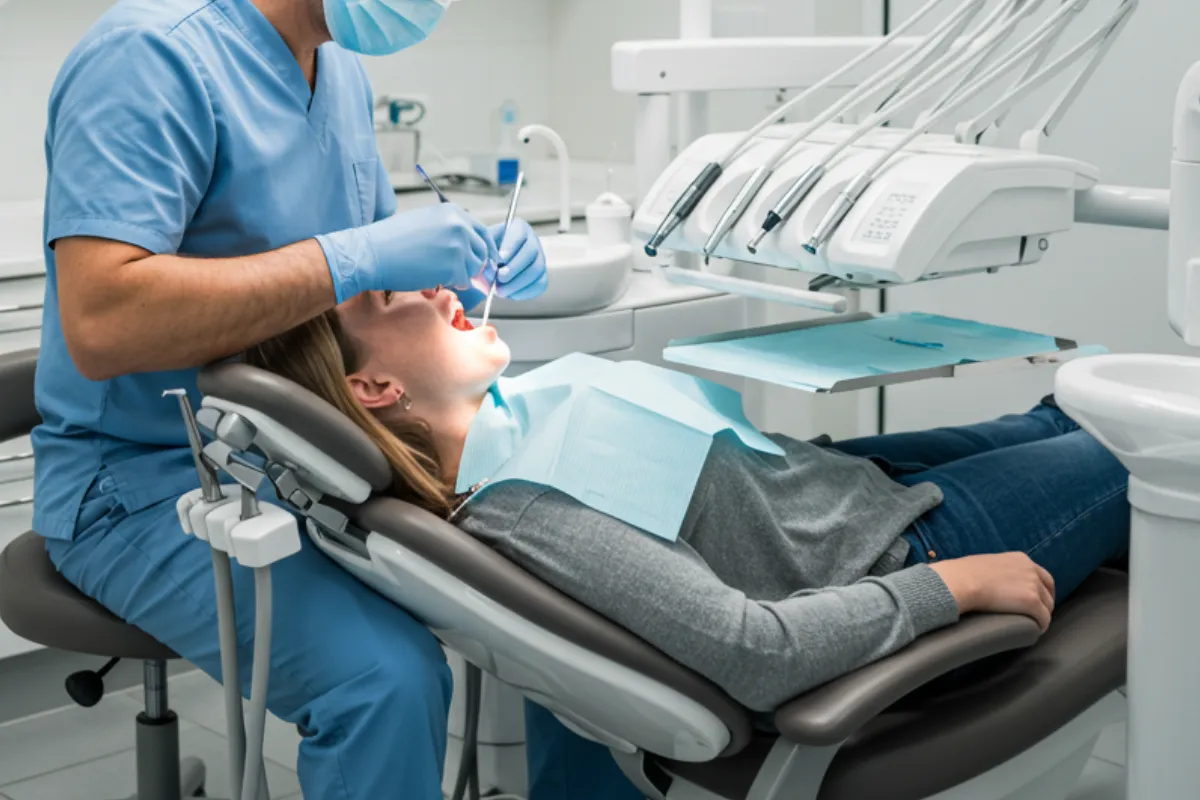
A dental assessment brings together several kinds of information. A dentist considers what can be seen, what imaging may show, your symptoms, and your likelihood of developing further decay.
Examining tooth surfaces and restorations
During an examination, the dentist looks at accessible tooth surfaces, grooves, and the edges of existing fillings or crowns. Cleaning and drying a surface can make subtle changes easier to see.
A tooth with a restoration still needs monitoring. A filling replaces damaged tooth structure, but it does not make the surrounding tooth immune to new decay. Plaque can collect near restoration margins, particularly where cleaning is difficult.
The dentist may also assess your bite, saliva, oral hygiene, and areas where food becomes trapped. Recent cavities, frequent snacking, dry mouth, and exposed root surfaces can all affect risk.
No single finding should be interpreted in isolation. A suspicious-looking spot needs context before a treatment decision is made.
Using X-rays when they are needed
Some surfaces cannot be examined directly, especially where neighboring teeth touch. Dental X-rays, when clinically indicated, can help reveal decay in these areas and sometimes around or beneath restorations.
Bitewing X-rays are commonly used to assess the spaces between back teeth. They can also help estimate how far a lesion extends through enamel and dentin.
X-rays have limits. They do not show every lesion, and some restorative materials can obscure nearby tooth structure. They also do not always establish whether a tooth surface has physically broken down.
The decision to take X-rays should reflect individual needs rather than a one-size-fits-all schedule. Your dentist considers your history, examination findings, previous images, and current risk. It is reasonable to ask what an image is intended to clarify and how the result could affect care.
Early Decay Does Not Always Need a Filling
One of the most useful distinctions in dental care is the difference between an early, noncavitated lesion and a cavity with actual surface breakdown.
In a noncavitated lesion, minerals have been lost, but the tooth surface remains intact. Depending on its location, depth, and activity, it may be possible to stop or reverse the process without drilling.
Management may include fluoride toothpaste, professionally applied fluoride, changes to eating habits, and better plaque removal. Some people may benefit from prescription-strength fluoride toothpaste when appropriate for their age and risk.
For selected lesions, a dentist may recommend a sealant or resin infiltration. These approaches are suitable in particular circumstances, not for every area of decay.
Monitoring is part of treatment, too. It involves reassessing the lesion and looking for evidence that it has stabilized or progressed. “We will monitor it” should come with an explanation of what you can do at home and when it needs another check.
An early lesion does not have to disappear visually to become inactive. A mark may remain after the decay process has stopped. The aim is a stable, healthy tooth, not necessarily a perfectly uniform color.
When Restorative Treatment Becomes More Likely
Once a cavity forms, the missing tooth structure does not grow back. Fluoride can help protect the remaining tooth, but it cannot rebuild a hole into its original shape.
Many cavitated lesions need restorative dentistry to remove or manage damaged tissue and restore a surface that can function and be cleaned. A filling may be appropriate when the damage is limited. A tooth with more extensive structural loss may need a different type of restoration.
Treatment also depends on whether the area is accessible for cleaning, how active the lesion is, and the person’s overall circumstances. In selected cases, methods such as silver diamine fluoride can help arrest decay, although they have limitations and can permanently darken the affected area.
If decay reaches the pulp and causes irreversible inflammation or infection, a filling alone may not resolve the problem. Root canal treatment, part of endodontics, may be needed to treat the inside of the tooth. A tooth that cannot be predictably restored may require extraction.
None of these outcomes can be predicted from pain alone. The treatment decision depends on the condition of the tooth.
Why Waiting for Pain Can Change Your Options
Consider two hypothetical situations.
In the first, a routine examination identifies an early lesion between two teeth. The surface is still intact. The person improves cleaning between the teeth, uses fluoride as advised, and reduces frequent sugary snacks. Follow-up assessments show that the lesion has stabilized.
In the second, someone delays assessment because the tooth feels fine. The lesion progresses and the surface breaks down. When it is eventually examined, a filling is needed.
These examples are not guarantees. Some lesions progress slowly, some remain inactive, and others advance more quickly. But earlier assessment generally gives you a better chance of having less invasive options.
Waiting for discomfort can also make timing harder. An appointment you could have scheduled calmly may become an urgent visit that disrupts sleep, work, or meals.
There is no need to feel embarrassed if it has been a long time since your last appointment. Dental anxiety, cost, access, and competing responsibilities are real barriers. The useful next step is an assessment of what is happening now, followed by a plan you understand.
Everyday Habits That Help Limit Decay
Preventing decay is largely about reducing repeated acid attacks and supporting the tooth’s ability to resist them. It does not require a perfect diet or an elaborate routine.
A few consistent habits make a meaningful difference:
Brush twice daily with fluoride toothpaste. Use an age-appropriate amount, and follow your dental professional’s advice about technique and fluoride strength.
Clean between teeth daily with floss or another suitable interdental cleaner. Choose a method you can use comfortably and consistently.
Reduce frequent exposure to sugary foods and drinks. Sipping a sweetened drink over several hours creates repeated opportunities for acid production.
Make water your usual drink between meals, and discuss persistent dry mouth with your dentist.
Attend examinations at an interval based on your individual risk and treatment needs.
Frequency matters alongside quantity. Eating something sweet with a meal is generally less harmful than repeatedly grazing on it throughout the afternoon.
Dry mouth deserves particular attention because saliva helps protect teeth. Medications, certain health conditions, and some medical treatments can reduce saliva. If your mouth often feels dry or sticky, mention it rather than assuming it is unavoidable. Do not stop a prescribed medication without speaking with the clinician who prescribed it.
Children Can Have Painless Decay, Too
Children may have decay without complaining, especially when lesions are early. They may also struggle to describe sensitivity or adapt by chewing on the other side.
Primary teeth matter even though they eventually fall out. They support eating and speech and help maintain space for permanent teeth. Untreated decay can lead to infection and affect a child’s comfort.
Pediatric dentistry includes assessing cavity risk, guiding home care, and considering preventive measures such as fluoride varnish or sealants. Caregivers should help with brushing until a child can clean thoroughly and reliably. Newly erupted permanent molars deserve attention because their grooves can be difficult to clean.
A child eating normally is encouraging. It is not a substitute for an examination.
When to Arrange an Appointment
Book a dental assessment if you notice a hole, a broken filling, repeated food trapping, new sensitivity, or a change in a tooth’s appearance. You should also arrange care if you are overdue for a recommended examination, even when everything feels comfortable.
Seek prompt dental care for persistent toothache, pain when biting, swelling, drainage, or a bad taste associated with a tooth. Severe pain that suddenly stops does not necessarily mean the underlying problem has resolved.
Facial swelling with fever, swelling near the eye, or difficulty swallowing needs urgent attention. Difficulty breathing is an emergency.
For a painless tooth, the message is less dramatic but still worth remembering: feeling fine and being healthy are not always the same. An examination can help distinguish a stable mark from active decay and an early lesion from damage that needs repair. That information gives you something more useful than waiting for pain: a chance to act while the options may still be simpler.